

Introducing MMU’s first industrial CT scanner – The Neoscan NXL
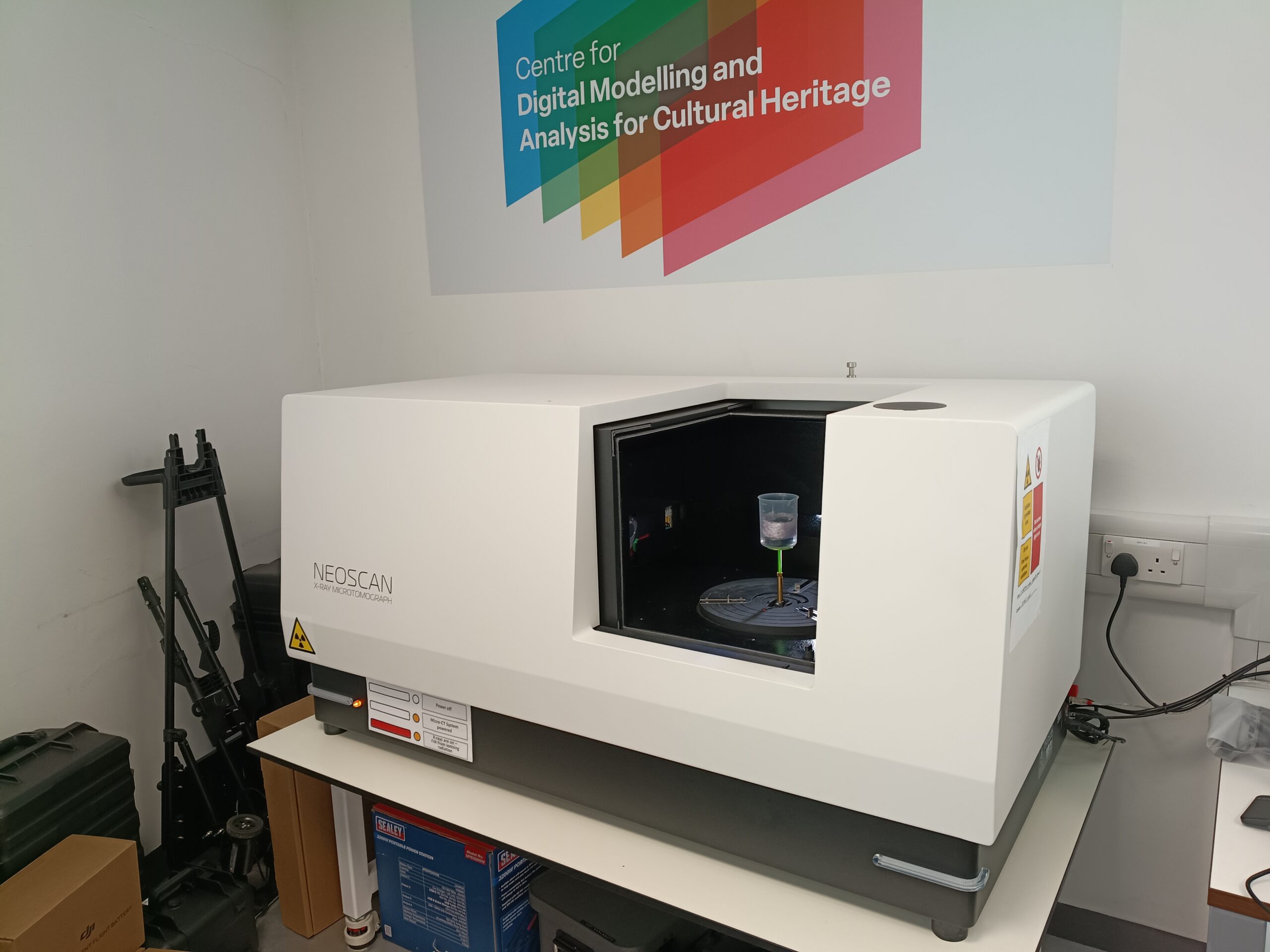
Manchester Metropolitan University's first industrial CT (computed tomography) scanner has been up and running for over month now. Purchased as part of the UKRI RICHeS grant used to set up the D-MACH lab, its primary application will be supporting research questions from Galleries, Libraries, Archives, and Museums (GLAM sector). However, we're opening access to researchers across MMU and wider, including external academics and companies, as well.
How does it work?
X-ray CT is a non-destructive testing method able to see inside solid object and provide a digital 3D dataset of the interior. It’s the same technology as medical CAT scanning only applied to non-living things.
It works by gathering X-ray images from all around an object. It uses this 2D data to interpret the interior position of parts of the object in 3D. This is possible because different materials absorb or transmit X-rays by different amounts. Bone blocks X-rays more than muscle and metal blocks X-rays more than bone. For a medical CT scan, this would mean that a CT scan of a hip replacement would show clearly the size, shape and location of the metal of the replacement, the patient’s bone and the surrounding muscle tissue.
CT scanners come in all different sizes and applications, so what is MMU’s scanner capable of? The original brief was a scanner which could image an object the size of a human skull and be versatile enough to image all manner of hand specimen sized museum artifacts. With a 160 kV, 75 W X-ray source it can image though wood, bone, pottery and even polymers with ease. The system can also image some metal components, but as with all techniques, the best way to know if something will work, is to try it!
What can it see?
The smaller the specimen, the higher the magnification that can be achieved, for example, here is a test scan of a foraminifera’s shell (a sea dwelling single celled organism). The foraminifera is 1.4 mm in diameter yet we can still make out the intricate texture of the shell.
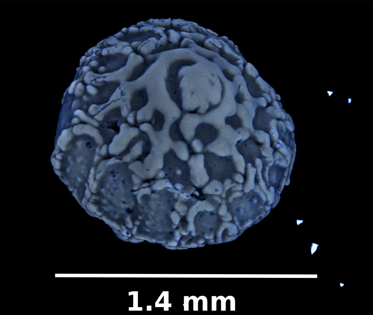
From testing, we are able to identify features as small as 5 microns (0.05 mm) on specimens 5 mm in diameter. For a typical hand specimen, like a small Roman ceramic oil lamp, we could resolve features within the lamp as small as a grain of sand. Remember, it can see differences in materials, that could be a maker’s mark, a crack inside the clay wall or another artifact hidden within.
CT can also give us measurable information about an object. For example:
- wall thickness of a container
- the amount of pore space in a rock
- how many separate objects or parts made up the whole sample
- true to life sizes for species comparison and determination
- quality assurance such as seeing if a 3D printed part matches its CAD model or contains defects not seen on the surface.
Part of the D-MACH workflow
Just like any 3D dataset, we can bring these data back into the real world via processes like 3D printing. This opens up the possibilities of interactive museum exhibits, tactile learning tools and in-hand data manipulation of something that would otherwise never be touched by human hands again, like a skull from a mummified animal! D-MACH’s location at MMU’s PrintCity means these tools are right on our doorstep.
We hope to combine our CT system with the other scanning equipment in the new D-MACH lab to create functional workflows to help curators, researchers, and curious members of the public explore collections like never before.

To speak to a member of the team with general questions visit here.
If you have a project in mind which might be suitable for the CT system, or any of the equipment at D-MACH, complete the form here.
